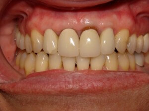
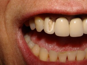
Here is a case of four ceramic dental crowns done on upper broken front teeth that we are quite proud of. The reason is not so much the appearance of the final result, but the workup. We hope that the workup (what you cannot see) will make the case last much longer.
Let us explain, with some history first:
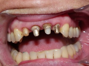
This is not the first time that these upper four front teeth have had crowns. In fact, the final result from the previous crowns looked pretty good! Four all- ceramic crowns were placed on the front teeth, after root canals and post/core buildups on three of them. (None of this done by Dr. Wong, in case you were wondering). The broken front teeth crowns came over several years.
This was completed under ten years ago, and functioned well until some slight marginal breakdown was noted on the lingual (inside) aspect of two of the crowns. Additionally, the margins at the front started showing the underlying darkness of the tooth roots.
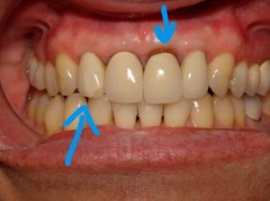
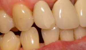
Finally, the crowns themselves started showing fracture lines and ultimately pieces broke off completely. This case was a cosmetic failure, and new dentistry was required.
The easy approach would have simply been to replace the crowns with new crowns just like the previous ones. However, stepping back and thinking does help the overall dental management.
1. Why did the crowns break?
This is key to the case. All-ceramic dental crowns do not break for no reason. There was no history of trauma to these crowns. If there were some defect in the manufacture of the crowns, they would have broken much sooner. There must have been biting forces exerted on the crowns that exceeded the strength of the ceramic material. Any dental treatment plan must include strategies to minimize these forces, or else a similar fate would likely befall the new crowns.
2. Are the underlying teeth sound?
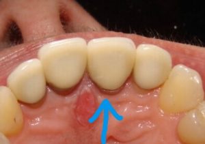
Fortunately, we were confident that the periodontal (gum) condition was satisfactory, despite the recession. Due to a smile line (the level that the upper lip rises to upon smiling) that did not expose the gumline, we opted not to alter any gingival levels via grafting. The overall gingival levels are not ideal, but due to patient goals and timeline, we opted against any alteration.
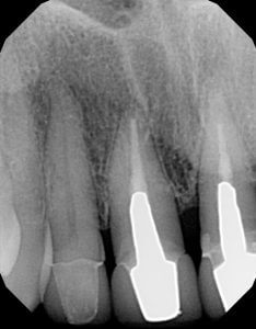
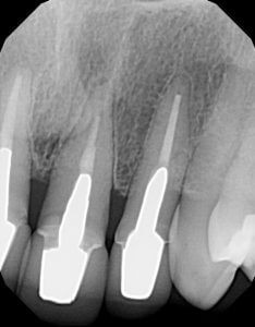
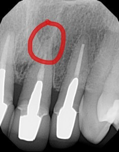
Still, we needed to know if the roots were acceptably healthy, and if the underlying teeth beneath the crowns were decay-free. The former would require x-rays, and the latter could be determined only by removal of the existing fractured crowns. As it turned out, one of the root canals was suspect, and required a referral to an endodontist for retreatment.
3. Are there any cosmetic deficiencies to the shapes of the old crowns that we wished to correct?
The midline required uprighting in order to be more vertical. Aside from this, the patient liked the shapes of the existing crowns, so we chose to keep them the same shape.
Management
Before preparing the teeth for the replacement crowns, we needed to:
1) Determine the proper bite position of the teeth.
Because of the fracture of front tooth crowns, we suspected that the patient actually had a Constricted Chewing Pattern. This means that upon chewing, the lower jaw actually needs more space anteriorly to close properly. Owing to the position of the upper crowns, the lower jaw was actually hammering away at the upper front teeth, and over the years, caused the ultimate weakening of the ceramics.
In order to determine the actual room required by the lower jaw to close properly, we gave the patient a Kois Deprogrammer to wear for two weeks. We confirmed deprogramming, and recorded the position on an articulator to facilitate lab communication.
Sure enough, the patient’s deprogrammed bite position indeed showed that it was heavy on the crowns.
2) Generate a wax simulation of the proposed final result
Using the articulator-mounted models, we asked the lab to provide us with a simulation of what they thought they could achieve, taking into account the previous constricted chewing envelope.
3) Referral to an endodontic specialist to retreat the root canal on the upper left front tooth. This was completed, and the patient was confirmed to be asymptomatic before proceeding with treatment. A new post and core assembly was constructed for that tooth as a new foundation for the new crown.
4) Preparation of the four front teeth
Although this is the “glamour” part of the procedures, this was relatively straightforward, and luckily did not reveal any further surprises in the form of large decay underneath the old crowns. The old crowns were removed and the teeth were prepared (shaved down) for new crowns. Standard impressioning with polyether impression material (Impregum) was used to capture a mould of the teeth, and a temporary set of crowns were made in he shape of the waxup teeth.
5) Refining of the temporary crowns
Before the lab was instructed to fabricate the final ceramic crowns, any adjustments to the bite or aesthetics of the crowns could be done to the temporary crowns. An impression of the temporary crowns was taken to show the lab what we really wanted.
5) Cementation of the new crowns
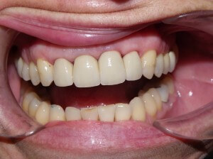
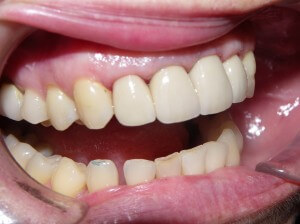
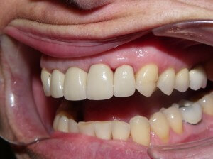
The final reveal! No problems with the fit! No adjustments were needed at all, and we thought the case looked great. More importantly, we are confident that the dentistry will last longer than they did the first time.
If you have concerns with the appearance of your front teeth, contact us! We will create a plan that will get you teeth that you can be proud to show off! We’d love to be your Barrie dentist.